
1/n TL;DR- Cell paper major claim re reprogramming ESC into 8-16cell stage "embryo founder cells EFCs" defined as 85-97% being OCT4+CDX2+GATA6+, is REFUTED by DATA in the SAME PAPER & PREPRINT. Immunostaining & scRNAseq prove CDX2-GATA6 rarely colocalize. https://www.cell.com/cell/full...

2/n Li et al. purported a concept of transiently capturing EFCs based on coexpression of OCT4/CDX2/GATA6 in 8-16cell embryos, after 60h of chemical treatment. Main proof shown is based on triple+ FACS intracellular staining for these genes (& NOT using triple reporter ESC)

3/n Intracellular FACS staining for TFs is a "dirty assay", each sample must have its OWN unstained and ISOTYPE MATCHED CONTROL to ensure true gating 4 positive staining. Wasn't done! & Preprint had GATING showing low%, was CHANGED in the Paper resulting in boost up to >85%!
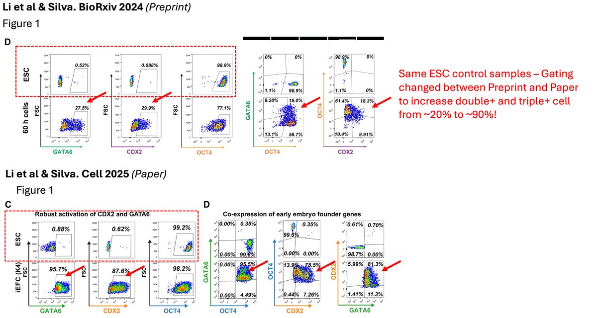
4/n why important? We need to distinguish btwn: 1)Naive ESC reverting back to 8-16 cell "EFCs"= most cells co-expressing OCT4/CDX2/GATA6 2)Naive ESC partially/on their way differentiating into TE or PrE = resulting in OCT4+ cells with CDX2+ or GATA6+, NOT CDX2/GATA6 co-expressed
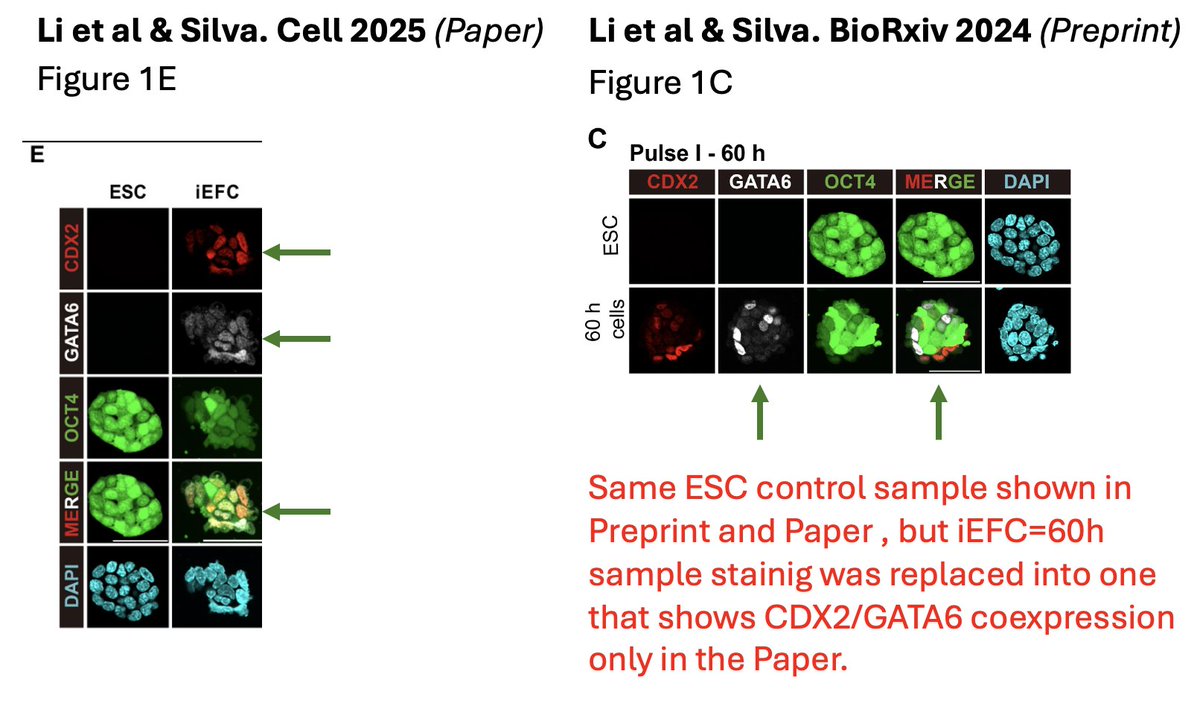
5/n why drown in intracellular stainings? Immunostaining for these markers r very easy to do in EFC=60h (names of same between Paper & Preprint https://tinyurl.com/tu6cv3cd). No quantitative & wideview stainings r shown. 2/3 iEFC stainings in Paper show no/rare CDX2+/GATA6+.

6/n Preprint: 3/3 iEFC stainings show no/rare CDX2+/GATA6+ (include the same 2 above) and one in Preprint Fig1C, which was replaced in Paper Fig 1E with one that shows fuzzy coexpression. In sum 3 out of 4 immunostainings shown, rare CDX2/GATA6 coexpression!
7/n Strangely straightforward scRNA-seq analysis of 60h=iEFC in Preprint showing rare CDX2/GATA6 coexpression & and that iEFC is basically a mix of Epi/PrE/TE, was NOT included in the published Paper although same dataset was used in both. & Nanog is reduced in TE-&PrE-like cells
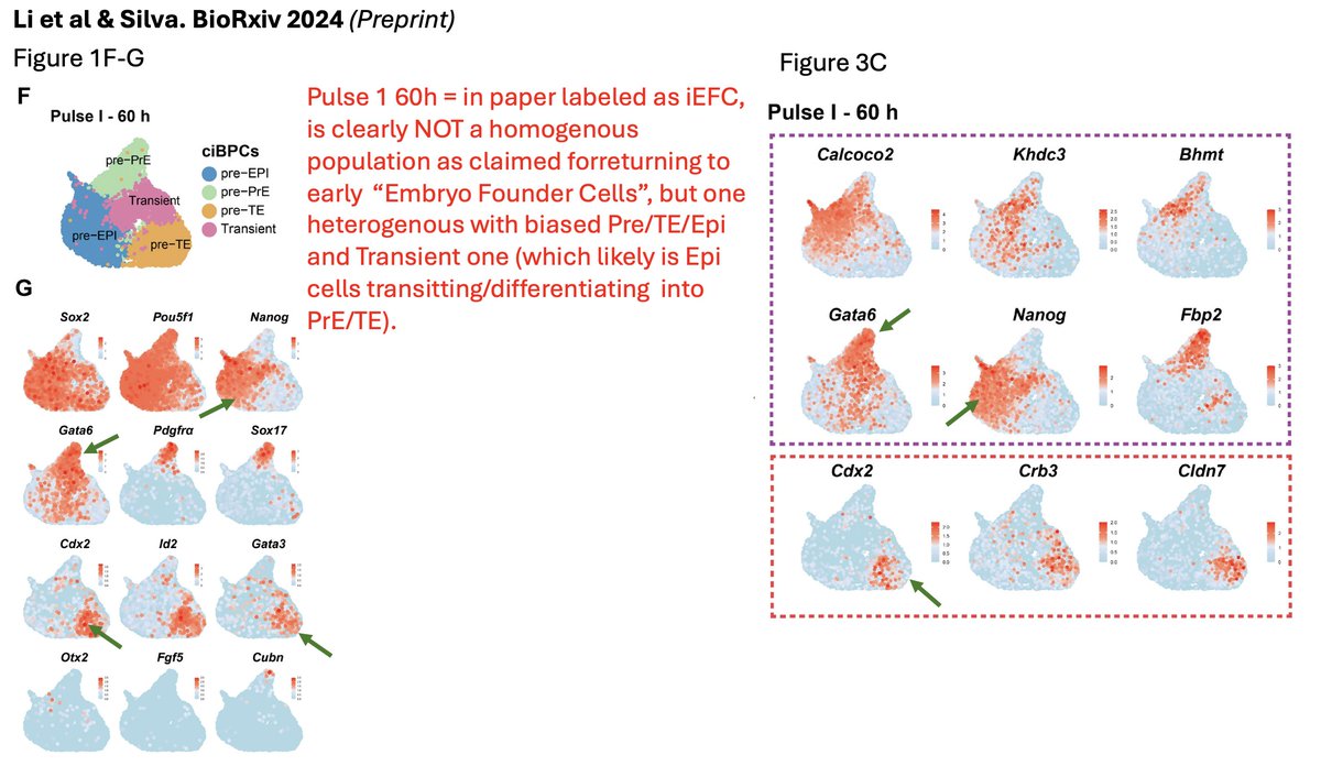
8/n Straightforward scRNA-seq analysis of 60h=iEFC in Preprint also showed cells are not going backwards in development, but forward. Gata6+ cells express Sox17 and Pdgfra which are not 8-16cell markers but of good old E4.5 PrE cells.

9/n some meta-analysis was done to claim iEFC fall on 8 cell. Upon close inspection - 8C and 32ICM are coloured similarly! and there is NO arrow pointing to 32ICM on the UMAP. it seems 32ICM cells are nicely overlapping with iEFCs? But again, simple scRNA-seq above says it all!

10/n in relation to the above concern, another panel of Paper in Fig.3F 32C ICM are marked (left) and according to this analysis on dataset used they also highly overlap with 16-Cell labeled in green? yet again why drown in scRNA-seq when immunostainings r so easy to solve this ?

11/n last one (plenty more but u should get the point by now) - iEFCs=60h in Preprint are shown to have a mix of PrE/Epi/TE , while in Paper they are homogeneously labeled in Magenta?

12/n That concludes our journal club on this. Preprints seem to be important to understand how data presentation can get so cherry picked & convoluted during review process to make bombastic claim (8cell EFC) no matter what. FACS gating info& quantitative immunostaining r a must!
Fin - in 2025 to make such a claim, relying on dubious intracellular FACS stainings should not be an acceptable standard. in the age of CRISPR - double or triple reporter ESCs should be a standard. Yet again - good old immunostaining in Paper Fig S1H-I tells us what "iEFCs" are!
"Zina" is one of my favorite songs, makes me think of my daughter Lina & now it takes a very different meaning >1500 Gaza children killed only since March when Israel broke ceasefire! Several were called Zina. This week another Zina died of starvation. https://www.youtube.com/watch?...