
1/ Shock isn’t “give fluids, then pressors, then inotropes.” That recipe misses the physiology. Here’s how to manage shock properly: 🧵 #MedX #haemodynamics
2/ First, rule out mechanical causes (tamponade, tension PTX, massive PE, acute valve rupture, etc). These need specific fixes.
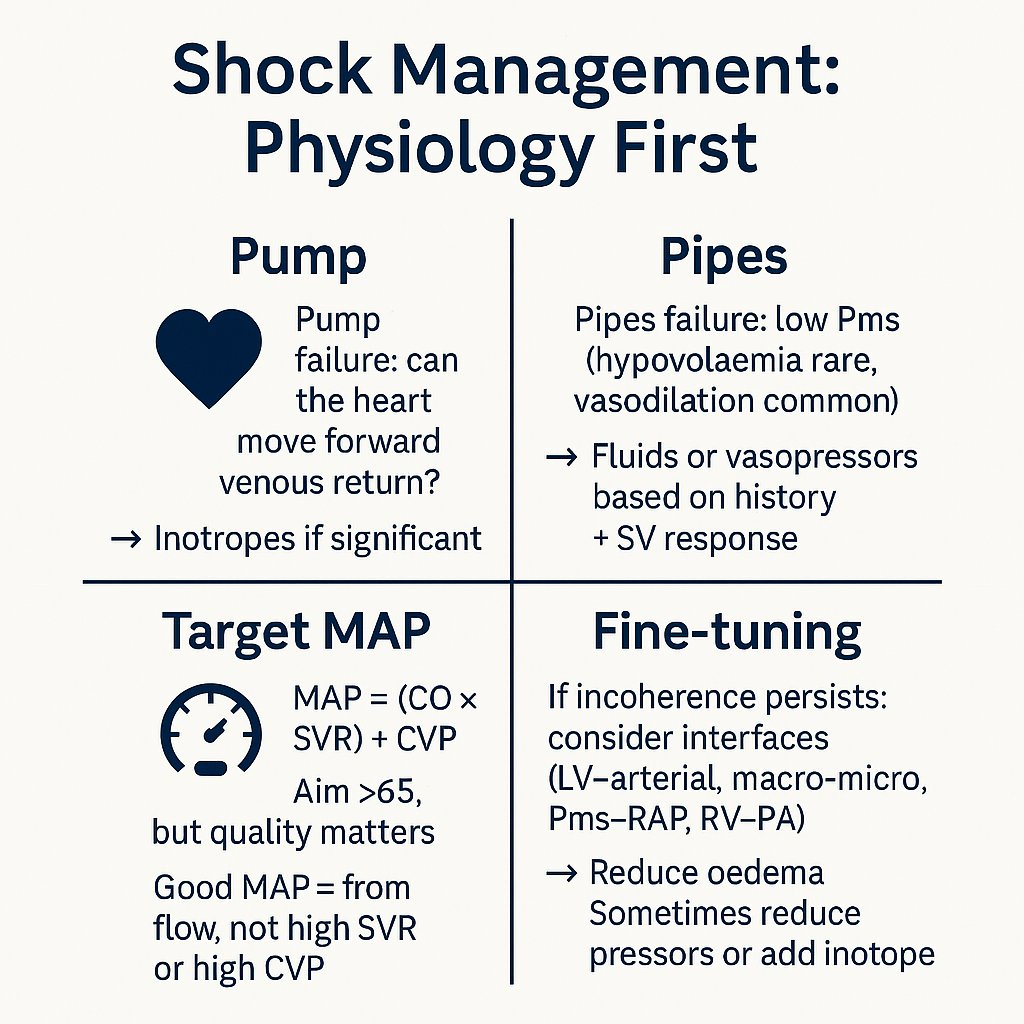
3/ Once you’ve ruled out mechanical causes, shock comes down to two mechanisms: • The pump fails (heart can’t move blood forward) • The pipes fail (low venular pressure → ↓mean systemic pressure (Pms) → ↓venous return) That’s the physiology behind almost every case.
4/ You can’t tell reliably from exam. Use history + echo; add a CO monitor if available. They save the hassle of repeating echo after every intervention — letting you track SV & SVR continuously. Sense-check uncalibrated monitors with echo once to be sure they’re in the right
5/ Pump failure ≠ automatic inotropes. The key is significance: Can the heart accept and move forward increased venous return if Pms rises? • If yes, dysfunction isn’t significant. • If no, it is — and that’s where therapy may help.
6/ Pipes physiology: A small, hyperdynamic LV is a common echo finding — but it’s not specific for low volume. It is usually vasodilation (low compliance, ↓Pms). True hypovolaemia is much less common. 💡 A common misconception: that vasodilation = high SV. In reality, untreated
7/ History separates causes: • GI loss/bleed/anaphylaxis → hypovolaemia • Sepsis/surgery/pancreatitis → vasodilation
8/ Fluid responsiveness ≠ hypovolaemia. It just means Pms-responsive. The neglected twin is vasopressor responsiveness — venoconstriction ↑stressed volume and Pms.
9/ Choose an intervention based on pre-test probability, then test the hypothesis. Measure SV before/after a small fluid, pressor, or inotrope change (VTI or a CO device). If SV doesn’t rise sustainably, rethink. 💡 A transient response to fluid means it wasn’t true hypovolaemia,
10/ Targets: don’t chase a CO number. Aim MAP >60–65, above the autoregulatory threshold for almost everyone. Peripheral perfusion is a practical bedside check you’re over the line.
11/ Not all MAP >60 is a good MAP. MAP = (CO × SVR) + CVP You want MAP generated by flow (CO), not by: – Excessive SVR (risk of surpassing critical closing pressure) – High CVP (venous congestion, shrinking the Pms–RAP gradient) 👉 See CCP threads for detail
12/ This links back to pump significance (Tweet 5). Inotropes: not “low EF = give dobutamine.” They only make sense if: – MAP ≥65 but SVR and/or CVP are high, or – MAP is low due to pump failure itself. In both cases, the heart cannot move forward extra venous return when Pms
13/ If macro looks right but the patient still looks poorly perfused → haemodynamic incoherence. This is where the interface model helps. https://x.com/icmteaching/stat...
14/ Interfaces for fine-tuning: • I1: LV → arterial coupling (LVEF/afterload) • I2: Macro → micro (MAP vs autoregulatory threshold, CCP) • I3: Capillary → venous (Pms–RAP gradient, congestion) • I4: RV → PA coupling (PH, ventricular interdependence) Occasionally, higher
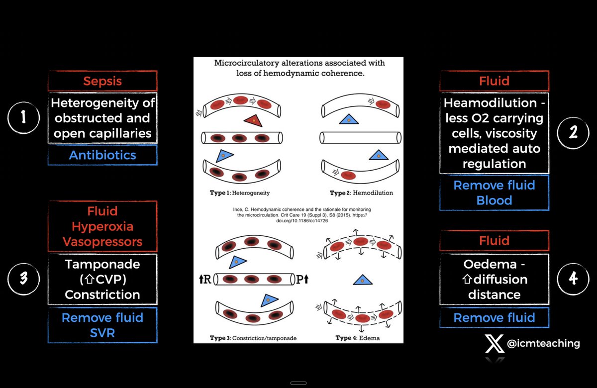
15/ Microcirculatory dysfunction (Ince): Heterogeneity (open/closed capillaries side by side) Haemodilution (low viscosity, less O₂ delivery) Tamponade/constriction (high CVP, CPP, impaired outflow) Oedema (↑diffusion distance) These explain why some patients remain incoherent
16/ Fine-tuning sometimes means going the other way. If SVR is very high, perfusion may be limited by critical closing pressure. Reducing vasopressors can improve flow — and sometimes inotropes help too, by allowing the heart to generate output at lower afterload.
17/ Case traps: • Oedematous, “fluid-responsive” patient improved with vasopressor + fluid removal (Interface 3 error). • Low EF but improved with pressor, not inotrope → example here 👉 https://x.com/icmteaching/stat... • “Hyperdynamic” echo after resus ≠ needs fluid. I’ll post case
18/ Key points: • Hypovolaemia is uncommon — don’t overfill • Vasodilation is common — squeeze • Treat pump failure only if significant • SV change is your bedside truth test • Reducing oedema almost always helps the microcirculation Start simple (pump vs pipes). If macro is
19/ Start here ⬇️ This is my framework for managing shock — simple to follow, physiologically robust, and practical at the bedside. https://x.com/icmteaching/stat...
@icmteaching @ThinkingCC @khaycock2 @msiuba @AndromedaShock @ArgaizR @edu_kattan @nickmmark @Wilkinsonjonny @NephroP @ross_prager This is great! You need a recipe book for each different scenario now!
@TheNarulaSeries @ThinkingCC @khaycock2 @msiuba @AndromedaShock @ArgaizR @edu_kattan @nickmmark @Wilkinsonjonny @NephroP @ross_prager Thank you 🙏🏼. The key is individualised therapy based on physiology. Not some illogical ‘one size fits all’ approach for a heterogeneous bundle of pathologies that cause shock. eg 30mls/kg of fluid 😬
@icmteaching @ThinkingCC @khaycock2 @msiuba @AndromedaShock @ArgaizR @edu_kattan @nickmmark @Wilkinsonjonny @NephroP @ross_prager Amazing trend!!!!! Respect! And Thank You!!!! 👍🏻🙂
@icmteaching @ThinkingCC @khaycock2 @msiuba @AndromedaShock @ArgaizR @edu_kattan @nickmmark @Wilkinsonjonny @NephroP @ross_prager Approach to Undifferentiated Circulatory Shock https://manualofmedicine.com/t...
@icmteaching @ThinkingCC @khaycock2 @msiuba @AndromedaShock @ArgaizR @edu_kattan @nickmmark @Wilkinsonjonny @NephroP @ross_prager This is a great description of the different underlying mechanisms that lead to shock. And each one Is treated differently from the next.
@icmteaching @ThinkingCC @khaycock2 @msiuba @AndromedaShock @ArgaizR @edu_kattan @nickmmark @Wilkinsonjonny @NephroP @ross_prager Unless we know the right markers for tissue hypo-perfusion better ….We tend to over treat SHOCK either with fluids, pressors or inotropes ! The myth lies with detection of tissue hypo-perfusion, NOT just the numbers of MAP, SBP or CVP Time for Organ Perfusion Pressure (OPP)!