
⭐️ Classic case: 60 y/o F presents w/ 3 years progressive pain in legs, difficulty with walking and balance, and occasional overflow incontinence. What is your diagnosis? #MedEd #medicine #Neurology #futureradres #radres #Neurosurgery #MRI @TheASNR #FOAMed


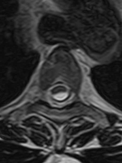
⭐️ Answer: Ventral cord herniation 🔷PATH: Acquired or congenital defect in the ventral dura allows the subarachnoid space and therefore the cord to slip through the defect into the epidural space
🔷CLINICAL: ▶️Chronic progressive myelopathy ▶️Progressive Brown Sequard syndrome can be seen if only one side of the cord herniates 🔷LOCATION: ▶️Ventral cord usually T2-T8 (normal kyphosis puts the cord in close proximity to the ventral dura)
🔷IMAGING: ▶️Morphology of the cord at the herniation is focally kinked (rather than the scalpel sign seen in a dorsal thoracic arachnoid web) ▶️No CSF between ventral cord and dura (find this one tough because it can look like this with webs too) ▶️ “Nuclear trail” sign


🔷DIFFERENTIAL: 1️⃣Dorsal thoracic arachnoid web: “Scalpel sign” rather than focal distortion/kinked appearance, may see CSF between cord and dura, may see band like interruption of CSF pulsation artifact at the level of the web (example in 🧵) 2️⃣Arachnoid cyst: Smooth
⭐️ Companion case of a dorsal thoracic arachnoid web with textbook scalpel sign 💡 Case courtesy of @MrFDA69


@MrFDA69 ⭐️ Another companion case of a dorsal thoracic arachnoid web with scalpel sign and focal band of interrupted CSF pulsation artifact


@MrFDA69 For more reading 📖 https://pubs.rsna.org/doi/abs/... https://radiopaedia.org/articl...
@daniel_gewolb @TheASNR Scalpel sign, The cord looks pulled anteriorly rather than pushed from posterior Ventral cord herniation
@daniel_gewolb @TheASNR Ventral cord herniation
@daniel_gewolb @TheASNR Ventral cord herniation
@daniel_gewolb @TheASNR Ventral cord herniation. Cracker case! Thankyou.
@daniel_gewolb @TheASNR Herniated cord
@daniel_gewolb @TheASNR Thoracic cord herniation due to dural defect
@daniel_gewolb @TheASNR Arachnoid web
@daniel_gewolb @TheASNR Ventral herniation
@daniel_gewolb @TheASNR One point This is a chronic thoracic myelopathy It should cause a suprasacral infrapontine bladder with high frequency, low volume urination and detrusor syphincter dyssenergia So there should be no overflow incontinence Overflow incontinence occurs when you have a
@daniel_gewolb @TheASNR Ventral cord herniation.
@daniel_gewolb @TheASNR Spinal cord herniation
@daniel_gewolb @TheASNR Ventral cord herniation
@daniel_gewolb @TheASNR Intra dural lesion
@daniel_gewolb @TheASNR Dorsal thorasic cord compression by IDEM- spinal arachnoid cyst( lesion has similar signal to csf)
@daniel_gewolb @TheASNR Dermoid tumor
@daniel_gewolb @TheASNR is it a Scapel sign?
@daniel_gewolb @TheASNR Nice case
@daniel_gewolb @TheASNR She’s pregnant